
Reflex Arc: Diagram and Components
Quick answer: A reflex arc is the fixed neural pathway, receptor → sensory (afferent) neuron → integration center (spinal cord) → motor (efferent) neuron → effector, that produces a rapid, involuntary response to a stimulus. Because the impulse is processed in the spinal cord or brainstem rather than the conscious brain, reflexes like the knee-jerk and the withdrawal from pain happen in milliseconds, protecting the body before damage occurs.
Key Facts: Reflex Arc
- A reflex action is a rapid, involuntary response to a stimulus; the reflex arc is the neural pathway (the wiring) that carries it out.
- The arc has 5 components in order: receptor → sensory (afferent) neuron → integration center → motor (efferent) neuron → effector.
- Sensory impulses enter the spinal cord through the dorsal root (cell bodies in the dorsal root ganglion); motor impulses leave through the ventral root (cell bodies in the ventral horn).
- The knee-jerk (patellar) reflex is monosynaptic: one synapse, sensory neuron directly onto motor neuron, no interneuron.
- The withdrawal reflex (snatching the hand from heat) is polysynaptic: it includes interneurons (relay neurons) between sensory and motor neurons.
- Spinal reflexes are integrated in the spinal cord; cranial reflexes (blinking, pupillary) in the brainstem; autonomic reflexes (blood-pressure control) run without conscious awareness.
- Doctors test reflexes clinically: an absent or exaggerated reflex reveals damage in the sensory or motor limb of the arc.
Touch a hot plate and your hand jerks back before you feel the pain. How is that possible? Because the message never went to your brain. It took a shortcut through the spinal cord: the reflex arc. This is the body's emergency hotline, and it follows the same five stations every single time. Learn the order once, and every reflex question becomes trivial.
What Is a Reflex Action? Action vs Arc
A reflex action is a rapid, automatic, involuntary response of the body to an external or internal stimulus. The reflex arc is the anatomical pathway, the fixed route the nerve impulse travels from the point of stimulation to the responding organ. Keep the two apart: action = the event (your hand jerking away); arc = the wiring (the five-component circuit). Examiners test this distinction directly.
The Five Components of the Reflex Arc (In Order)
- Receptor: a specialised sensory structure that detects the stimulus, e.g. pain receptors in the skin or stretch receptors (muscle spindles) in a muscle.
- Sensory (afferent) neuron: carries the impulse toward the central nervous system.
- Its cell body sits in the dorsal root ganglion, a swelling on the dorsal root just outside the spinal cord.
- Its axon enters the spinal cord through the dorsal root.
- Integration center: the gray matter of the spinal cord (or the brainstem for cranial reflexes), where the impulse is processed.
- In polysynaptic reflexes this includes one or more interneurons (relay neurons) linking sensory to motor neurons.
- In monosynaptic reflexes the sensory neuron synapses directly on the motor neuron, with no interneuron.
- Motor (efferent) neuron: carries the impulse away from the CNS to the effector.
- Its cell body sits in the ventral horn of the spinal cord's gray matter.
- Its axon leaves the spinal cord through the ventral root.
- Effector: the muscle or gland that produces the response, e.g. the biceps contracting to withdraw the hand.
Between every two neurons lies a tiny gap, the synapse, across which the impulse passes chemically via neurotransmitters. Memory hook: sensory goes in (dorsal root, afferent = arriving), motor goes out (ventral root, efferent = exiting).
Labeled Diagram of the Reflex Arc (Guide)
A standard labeled diagram of a spinal reflex arc shows the spinal cord in cross-section with the nerve pathway traced through it. Label these parts:
| Label | Part | What it does |
|---|---|---|
| A | Receptor | Detects the stimulus: heat, pressure, pain, stretch |
| B | Sensory (afferent) neuron | Carries the impulse from the receptor toward the CNS via the dorsal root |
| C | Dorsal root ganglion | Houses the sensory neuron's cell body |
| D | Integration center | Gray matter of the spinal cord, where interneurons (in polysynaptic arcs) process the impulse |
| E | Motor (efferent) neuron | Carries the impulse out via the ventral root; cell body in the ventral horn |
| F | Effector | Muscle or gland that produces the response |

How the Reflex Arc Works: Step by Step
- Detection: a stimulus (e.g. a pinprick) activates pain receptors in the skin.
- Transmission inward: the sensory neuron fires; the impulse travels along its axon toward the spinal cord through the dorsal root. (At the molecular level, this impulse is an electrical wave driven by Na+ and K+ ions moving across the axon membrane, the ion transport explained in our article on transport across the cell membrane.)
- Processing: inside the spinal cord's gray matter, the sensory neuron synapses with an interneuron, which passes the signal to the motor neuron. A branch of the signal also climbs to the brain, which is why you feel the pain a moment after your hand already moved.
- Transmission outward: the motor neuron carries the impulse out of the spinal cord through the ventral root to the effector.
- Response: the muscle contracts and the hand is withdrawn, all within a fraction of a second, with no conscious decision.
Worked Example 1: The Knee-Jerk (Patellar) Reflex
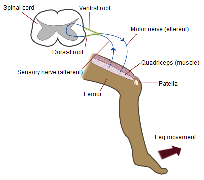
The patellar reflex is the textbook monosynaptic reflex, the one doctors test with a tap below the kneecap:
- The tap stretches the patellar tendon → stretch receptors in the quadriceps detect the stretch.
- The sensory neuron fires → the impulse enters the spinal cord via the dorsal root.
- One synapse directly onto the motor neuron: no interneuron, hence monosynaptic.
- The motor neuron fires → the impulse leaves via the ventral root.
- The quadriceps contracts → the lower leg kicks forward, all in about 50 milliseconds, with no brain involvement.
Worked Example 2: The Withdrawal Reflex
Touching a hot object triggers the withdrawal (flexor) reflex, the classic polysynaptic example:
- Heat activates pain receptors in the skin of the hand.
- Sensory neurons carry the signal into the spinal cord via the dorsal root.
- Interneurons in the gray matter relay the signal: they excite the flexor motor neurons (biceps contracts, hand pulled back) while simultaneously inhibiting the extensor motor neurons (triceps relaxes), so the two muscles do not fight each other.
- A second set of interneurons crosses to the opposite side of the cord and triggers the crossed-extensor reflex, stiffening the other leg so you do not fall over.
- The hand is withdrawn in milliseconds; the brain receives a delayed copy and you feel the burn afterwards.
Monosynaptic vs Polysynaptic Reflexes
| Feature | Monosynaptic | Polysynaptic |
|---|---|---|
| Synapses in the CNS | One (sensory → motor directly) | Two or more (interneurons included) |
| Neurons involved | 2 | 3 or more |
| Speed | Fastest reflex pathway | Slightly slower |
| Examples | Knee-jerk (patellar), Achilles (ankle-jerk) | Withdrawal from pain, crossed-extensor, pupillary |
Types of Reflexes: Spinal, Cranial and Autonomic
Reflexes are also classified by where they are integrated:
| Type | Integration center | Examples |
|---|---|---|
| Spinal reflexes | Spinal cord | Knee-jerk, Achilles reflex, withdrawal from pain, crossed-extensor |
| Cranial reflexes | Brainstem | Blinking, pupillary light reflex, corneal reflex, coughing, sneezing (protective reflexes of the airways; see the mechanism of breathing in man) |
| Autonomic reflexes | Spinal cord / brainstem (involuntary) | Baroreceptor reflex controlling blood pressure, pupillary dilation in dim light, salivation |
| Conditioned (acquired) reflexes | Brain (learned) | Salivating at the smell of food, Pavlov's dogs |
| Unconditioned (inborn) reflexes | Spinal cord / brainstem | Sucking in newborns, grasping, blinking |
The principle is identical in every case: a short arc that bypasses conscious thought, trading deliberation for speed.
Why Are Reflexes Important?
- Protection: instant responses (withdrawal from pain, blinking) act before tissue damage occurs.
- Posture and balance: stretch reflexes keep muscles at the right tension for standing upright and moving smoothly.
- Clinical testing: doctors test reflexes (knee-jerk, ankle-jerk, Babinski) to check the nervous system: an absent reflex suggests a lesion in the arc, an exaggerated one suggests damage higher up.
- Coordination: nervous reflexes handle the split-second work, while hormones handle slower, long-term regulation: the two great coordination systems of the body.
The #1 trap: students write that the impulse goes to the brain. In a spinal reflex it goes to the spinal cord; the brain only gets a delayed copy. The second trap: mixing up the dorsal root (sensory, in) with the ventral root (motor, out). When asked to "draw and label the reflex arc", draw the spinal cord in cross-section, trace the impulse with arrows, and label all five components plus the synapse: that diagram alone can earn full marks.
Frequently Asked Questions
Why is a reflex faster than a voluntary action?
The impulse travels a shorter path, receptor → spinal cord → effector, skipping conscious processing in the higher brain centers. Fewer synapses, fewer decision steps and a shorter distance mean a response in milliseconds instead of tenths of a second.
What is the difference between a reflex action and a reflex arc?
The reflex action is the event (your hand jerking away). The reflex arc is the neural pathway (the five-component circuit) that carries it out. Action = what happens; arc = the wiring.
Where is the cell body of the sensory neuron?
In the dorsal root ganglion, a swelling on the dorsal root just outside the spinal cord. The motor neuron's cell body, by contrast, sits in the ventral horn of the spinal cord's gray matter.
Are all reflexes spinal? What about blinking?
Most fast protective limb reflexes are spinal (withdrawal, knee-jerk). But some are cranial: blinking, the pupillary light reflex, coughing and sneezing are integrated in the brainstem. The principle is identical: a short arc bypassing conscious thought.
What happens if the reflex arc is broken, say the motor neuron is cut?
The reflex disappears: the effector never receives the message. This is exactly how doctors test for nerve damage, a missing knee-jerk can reveal a lesion in the sensory or motor limb of the arc.
Is the knee-jerk reflex present in newborns?
Yes, and newborn reflex testing goes further: babies show inborn reflexes like sucking, rooting and grasping that disappear as the brain matures. Their absence or persistence past the expected age helps paediatricians detect developmental problems.
Practice MCQs
Q1. The correct sequence of components in a reflex arc is:
- Receptor → motor neuron → spinal cord → sensory neuron → effector
- Receptor → sensory neuron → spinal cord → motor neuron → effector
- Stimulus → motor neuron → receptor → sensory neuron → response
- Effector → sensory neuron → spinal cord → motor neuron → receptor
Answer: B — The impulse travels from receptor through the sensory neuron to the CNS, then out via the motor neuron to the effector.
Q2. The knee-jerk (patellar) reflex is an example of a:
- Polysynaptic reflex
- Monosynaptic reflex
- Cranial reflex
- Conditioned reflex
Answer: B — The sensory neuron synapses directly on the motor neuron in the spinal cord, with no interneuron between them.
Q3. The gap between two neurons in a reflex arc is called the:
- Node of Ranvier
- Axon hillock
- Synapse
- Myelin sheath
Answer: C — The impulse crosses the synaptic gap chemically via neurotransmitters.
Q4. In a reflex arc, the cell body of the sensory neuron is located in the:
- Ventral horn
- Dorsal root ganglion
- Grey matter of the spinal cord
- Effector muscle
Answer: B — Sensory neuron cell bodies lie in the dorsal root ganglion, just outside the spinal cord.
Q5. Which of the following is a cranial reflex?
- Knee-jerk reflex
- Withdrawal reflex
- Pupillary light reflex
- Ankle-jerk reflex
Answer: C — The pupillary light reflex is integrated in the brainstem (a cranial reflex); the others are spinal reflexes.
Q6. Afferent (sensory) neurons carry impulses:
- From the CNS to the effector
- From the receptor to the CNS
- From muscle to muscle
- Within the brain only
Answer: B — Afferent means "carrying toward": sensory neurons bring signals from receptors into the CNS; efferent (motor) neurons carry orders out.
Q7. The withdrawal reflex (snatching the hand from a hot object) is a:
- Monosynaptic reflex
- Polysynaptic reflex
- Cranial reflex
- Conditioned reflex
Answer: B — Withdrawal involves interneurons between sensory and motor neurons, so it is polysynaptic (unlike the monosynaptic knee-jerk).
Q8. The effector in a reflex arc is the:
- Receptor detecting the stimulus
- Sensory neuron
- Muscle or gland that carries out the response
- Spinal cord
Answer: C — The impulse crosses the synaptic gap chemically via neurotransmitters.
Q9. A conditioned reflex differs from an unconditioned (inborn) reflex in being:
- Learned through experience and association
- Present at birth
- Faster than any inborn reflex
- Always spinal
Answer: A — Conditioned reflexes (e.g. salivation at a bell) are acquired by learning; unconditioned reflexes are born.
Q10. Doctors test reflexes like the knee-jerk during a clinical examination to assess:
- Muscle strength alone
- The functional integrity of the reflex pathway and nervous system
- Blood pressure
- Vision
Answer: B — Absent or exaggerated reflexes reveal damage or disease in the sensory/motor nerves or spinal cord segments.
Q11. Motor impulses leave the spinal cord through the:
- Dorsal root
- Ventral root
- Central canal
- Dorsal column
Answer: B — Ventral roots carry efferent (motor) fibres to muscles and glands.
Q12. Which of the following is NOT part of a reflex arc?
- Receptor
- Interneuron
- Effector
- Cerebrum as a voluntary decision centre
Answer: D — A reflex arc bypasses conscious decision-making; the cerebrum is not part of the arc.
Short Questions
Q1. What is a reflex arc? Name its five components in order. See answer
A reflex arc is the fixed neural pathway that carries the impulse responsible for an automatic, involuntary response (a reflex action). Its five components, in strict order, are: (1) the receptor that detects the stimulus, (2) the sensory (afferent) neuron that carries the impulse toward the CNS, (3) the integration center in the spinal cord or brainstem, (4) the motor (efferent) neuron that carries the impulse outward, and (5) the effector (muscle or gland) that produces the response.
Do not confuse the reflex action with the reflex arc: the action is the event (the hand jerking away), while the arc is the wiring. Examiners test this distinction directly, often as a one-mark definition pair.
Between every two neurons lies the synapse, the tiny gap across which the impulse passes chemically via neurotransmitters. The full chain is therefore: stimulus → receptor → sensory neuron → (interneuron) → motor neuron → effector → response.
Q2. Why is a reflex action faster than a voluntary action? See answer
A reflex is faster because the impulse travels only as far as the spinal cord (or brainstem) and never waits for conscious processing in the higher brain centers. The route is shorter, the synapses fewer, and no decision-making step intervenes, so the response arrives in milliseconds.
A voluntary action, by contrast, must travel up to the cerebrum, be interpreted, decided upon, and sent back down, a round trip through many more synapses that takes tenths of a second. That delay is the difference between touching a hot plate and being burned by it.
The speed is protective by design: reflexes guard the body against injury and maintain essential functions such as posture, balance and pupil size without burdening conscious thought. The nerve impulse itself is an electrical wave driven by Na+ and K+ crossing the axon membrane, the ion transport explained in our article on transport across the cell membrane.
Q3. Differentiate between monosynaptic and polysynaptic reflexes with examples. See answer
A monosynaptic reflex has a single synapse in the CNS: the sensory neuron connects directly to the motor neuron, with no interneuron. Only two neurons are involved, making it the fastest reflex pathway. The classic example is the knee-jerk (patellar) reflex, and the Achilles (ankle-jerk) reflex works the same way.
A polysynaptic reflex has two or more synapses: one or more interneurons (relay neurons) sit between the sensory and motor neurons, so three or more neurons participate. It is slightly slower but far more flexible. The classic example is the withdrawal reflex (snatching the hand from heat), which also recruits the crossed-extensor reflex to keep you upright.
Most reflexes in the body are polysynaptic; only a few stretch reflexes like the knee-jerk are monosynaptic. Examiners love the trap of calling the withdrawal reflex monosynaptic: remember that anything involving pain and coordinated muscle groups needs interneurons.
Q4. What is the difference between afferent and efferent neurons? Name their roots. See answer
A afferent (sensory) neurons carry impulses toward the central nervous system, from the receptors inward. Their cell bodies lie in the dorsal root ganglion, and their axons enter the spinal cord through the dorsal root. Think: afferent = arriving, dorsal = sensory, in.
Efferent (motor) neurons carry impulses away from the CNS, outward to the effector muscles and glands. Their cell bodies lie in the ventral horn of the spinal cord's gray matter, and their axons leave through the ventral root. Think: efferent = exiting, ventral = motor, out.
The dorsal/ventral root mix-up is one of the commonest exam errors: students write "motor impulses enter through the dorsal root". The memory hook "sensory in, motor out" (dorsal in, ventral out) prevents it.
Q5. Describe the knee-jerk reflex as a worked example of a monosynaptic arc. See answer
The doctor taps the patellar tendon below the kneecap, stretching the quadriceps muscle. Stretch receptors (muscle spindles) in the quadriceps detect this stretch and fire the sensory neuron, whose impulse enters the spinal cord through the dorsal root.
Inside the gray matter, the sensory neuron synapses directly onto the motor neuron: this single synapse, with no interneuron, is what makes the reflex monosynaptic. The motor neuron fires, and its impulse leaves the cord through the ventral root to the quadriceps.
The quadriceps contracts and the lower leg kicks forward, all in about 50 milliseconds, with no brain involvement. Meanwhile a copy of the signal climbs to the brain, which is why you feel the tap a moment after your leg already kicked.
Q6. Why do doctors test reflexes? What do abnormal results mean? See answer
Doctors test reflexes such as the knee-jerk, ankle-jerk and Babinski responses to check the functional integrity of the nervous system. Each reflex probes one complete arc: receptor, sensory nerve, spinal cord segment, motor nerve and muscle. It is one of the quickest neurological examinations available.
An absent or diminished reflex suggests a lesion in the arc itself: damage to the sensory nerve, the spinal cord segment, the motor nerve, or the muscle. A classic example is peripheral neuropathy, where the ankle-jerk disappears early.
An exaggerated (hyperactive) reflex suggests the opposite: damage higher up, in the brain or descending tracts, which normally dampen spinal reflexes. So the pattern of normal, absent and exaggerated reflexes maps the site of neurological disease, which is why the reflex hammer is a standard part of every neurological examination.
References
- OpenStax, Anatomy & Physiology 2e, §12.6 "Nervous System Disorders" and §13.4 "The Spinal Cord": OpenStax A&P 2e — the spinal cord and reflexes.
- Hickman, C.P., Roberts, L.S., Keen, S.L. et al. Integrated Principles of Zoology. McGraw-Hill Education. (Nervous coordination; reflex arcs.)
- Miller, S.A. & Harley, J.P. Zoology. McGraw-Hill. (Control and coordination in animals.)
- NCBI Bookshelf, StatPearls: "Monosynaptic Reflex": NCBI Bookshelf — monosynaptic reflex physiology.
Comments
Leave a comment